How a MedTech Startup Used AI to Improve Diagnostic Accuracy by 22%: A Paige AI Case Study offers a practical approach for teams looking to improve efficiency and outcomes.
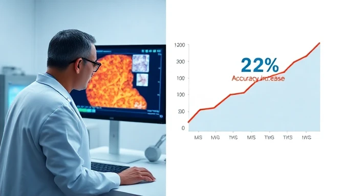
Paige AI's computational pathology platform helped MedTech startup, Histoview Diagnostics, achieve a 22% improvement in diagnostic accuracy for complex cancer cases, demonstrating a significant leap beyond traditional methods. This outcome wasn't achieved overnight; it required a strategic blend of advanced AI, meticulous data integration, and a phased implementation approach. For healthcare professionals navigating the evolving landscape of diagnostic tools, understanding this ai diagnostics case study offers a blueprint for leveraging AI to enhance precision and patient outcomes in 2026 and beyond.
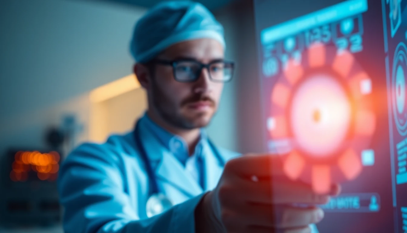
Meet Dr. Anya Sharma: Pathologist's Pursuit of Precision

Dr. Anya Sharma, co-founder and Chief Medical Officer of Histoview Diagnostics, recognized the critical need for greater precision in histopathology. Her MedTech startup, established in 2023, aimed to provide specialized diagnostic services for oncologists, focusing on challenging solid tumor cases where subtle morphological patterns often dictate treatment pathways. The core of Histoview's mission was to reduce diagnostic discrepancies and improve turnaround times, ensuring patients received accurate prognoses faster. Dr. Sharma, a board-certified pathologist with 15 years of experience in oncologic pathology, understood the inherent limitations of human visual analysis, especially under increasing caseload pressures. She believed that AI could augment, rather than replace, human expertise, pushing the boundaries of what was diagnostically possible. Her team comprised 12 highly skilled pathologists, 8 histotechnologists, and a small but agile software development unit, all operating under strict CLIA and CAP guidelines.
The Pre-AI Diagnostic Bottleneck: Before Paige AI
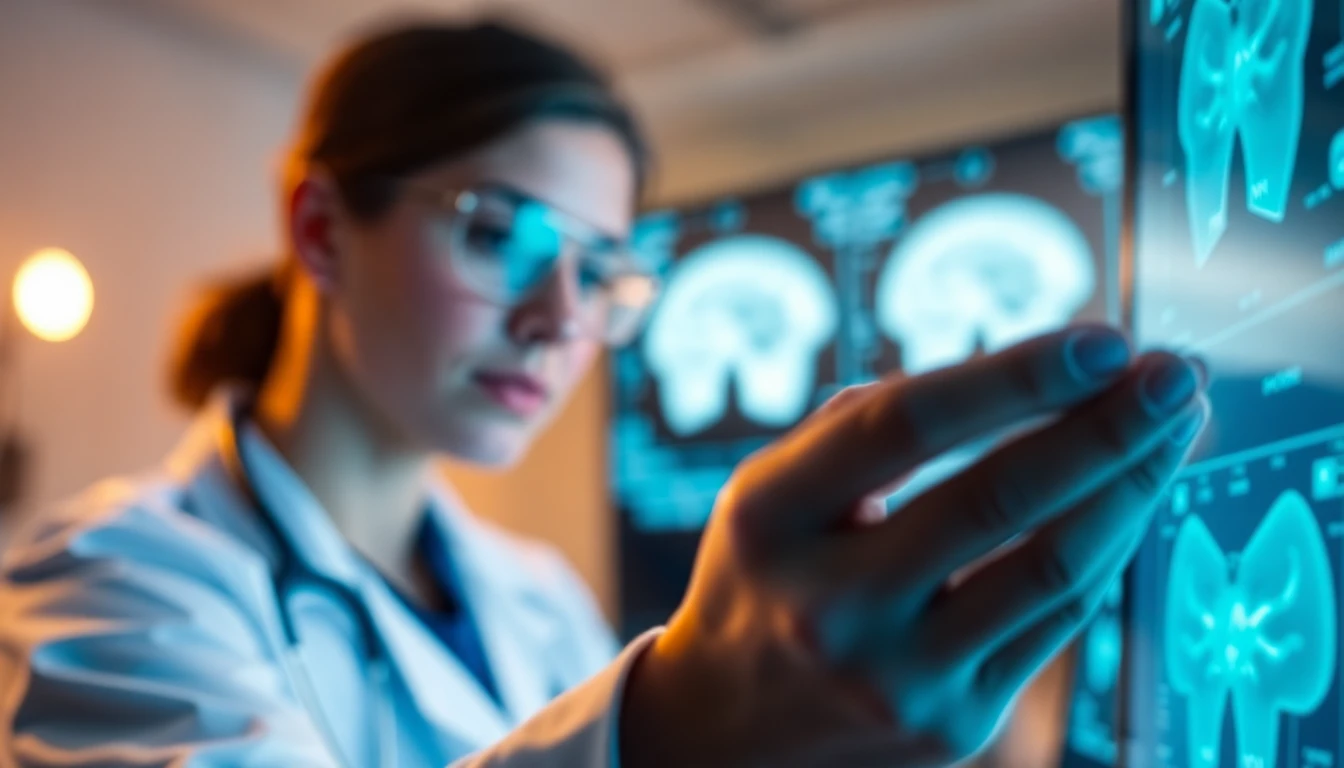
Before integrating AI, Histoview Diagnostics relied on a fully digital pathology workflow, utilizing high-resolution whole-slide imaging (WSI) and a robust PACS (Picture Archiving and Communication System) for image management. While this eliminated glass slides and streamlined remote review, the diagnostic process itself remained heavily dependent on individual pathologist interpretation. This human-centric model, though expert-driven, presented several inherent challenges that impacted accuracy and efficiency. Complex cases, particularly those involving heterogeneous tumor microenvironments or subtle invasion patterns, often required multiple pathologist reviews, extensive immunohistochemistry (IHC) staining, and sometimes even external consultations, prolonging the diagnostic cycle.
Manual Review: Baseline Error Rates
Histoview's internal audits, conducted quarterly, revealed a baseline diagnostic discrepancy rate of approximately 4.5% for their most challenging cases, defined as those requiring secondary review or resulting in a modified diagnosis after additional testing. This metric, while lower than industry averages for general pathology, was still a significant concern for a startup specializing in high-stakes oncology. These discrepancies often led to delayed treatment initiation, additional invasive procedures, and increased emotional burden for patients. Furthermore, the average time to definitive diagnosis for these complex cases stood at 7.2 days, including all ancillary studies and consultations. The human factor, characterized by fatigue, cognitive load, and the sheer volume of data in a single whole-slide image (often gigapixel-sized), contributed to these rates. Pathologists spent considerable time manually annotating regions of interest, meticulously counting mitotic figures, or visually estimating tumor burden, all tasks prone to inter-observer variability.
Prior Digital Pathology Gaps
Histoview Diagnostics had already invested heavily in a state-of-the-art digital pathology infrastructure, including a Philips IntelliSite Pathology Solution for scanning and a Sectra PACS for image storage and viewing. These systems improved workflow efficiency by digitizing slides and enabling remote access, but they did not inherently enhance diagnostic interpretation. The tools available within these platforms primarily focused on image navigation, basic measurement, and annotation—functions that digitized existing manual processes rather than fundamentally transforming them. Dr. Sharma's team experimented with early-stage image analysis software that could quantify specific stains or measure tumor size, but these tools lacked the sophisticated pattern recognition capabilities required for nuanced morphological assessment. They often produced high false-positive rates, necessitating extensive manual validation, which negated any potential time savings. The integration of these disparate tools was also cumbersome, requiring significant IT overhead and often leading to data silos, making a truly seamless diagnostic ecosystem elusive.
⚠️ Caution: Implementing digital pathology alone does not automatically improve diagnostic accuracy. Without integrated AI, it primarily digitizes existing manual workflows, potentially exposing the limitations of human visual analysis on a larger scale.
The Paige AI Solution Stack: Precision Pathology Platform
After extensive research and pilot testing with several AI pathology platforms, Histoview Diagnostics selected Paige AI, specifically its Paige Prostate and Paige Breast modules, complemented by the foundational FullFocus platform. Paige AI, as of 2026, is a leading computational pathology company focused on applying artificial intelligence to cancer diagnostics. Their platform is built on deep learning models trained on massive datasets of digitized pathology slides, annotated by expert pathologists. This extensive training allows the AI to identify subtle morphological features and patterns that are indicative of disease, often with a level of consistency and speed unmatched by human review alone.
Core AI Modules and Data Integration
Histoview Diagnostics deployed the following Paige AI modules:
- Paige FullFocus (Version 3.1, as of 2026): This foundational platform provides a universal viewer for whole-slide images and serves as the central hub for AI integration. It handles image loading, navigation, and annotation, ensuring compatibility with Histoview's existing Philips WSI scanners and Sectra PACS. FullFocus offers robust API support for integrating with laboratory information systems (LIS) like Epic Beaker, facilitating automated case routing and results reporting. Its cloud-native architecture (AWS-hosted) ensures scalability and data security, crucial for HIPAA compliance.
- Paige Prostate (Version 4.0, as of 2026): An FDA-cleared (in the US, specific regulatory clearances vary by region) AI solution for detecting prostate cancer, Gleason grading, and quantifying tumor burden. It
automatesthe identification of suspicious regions,quantifiestumor percentages, andsuggestsGleason patterns, providing pathologists with objective, reproducible metrics. - Paige Breast (Version 2.5, as of 2026): This module
assistsin the detection of breast cancer andclassifieskey features such as tumor type and grade. It is particularly adept at identifying micrometastases in lymph nodes and assessing tumor cellularity, often challenging tasks for manual review.
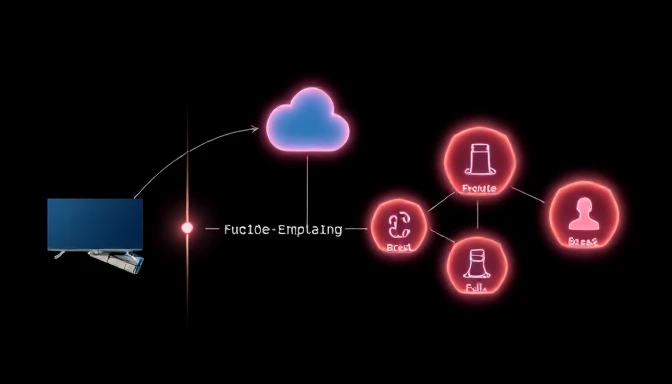
Data integration was a critical step. Histoview Diagnostics configured a secure, bidirectional interface between their Sectra PACS and Paige FullFocus using DICOMweb standards. This allowed for automated ingestion of newly scanned WSIs into the Paige platform for AI analysis and the seamless return of AI-generated insights (e.g., heatmaps, annotations, quantitative scores) back into the PACS for pathologist review. The LIS was also integrated to pull patient demographics and order information, ensuring the AI analysis was contextually relevant. This ships an n8n integration that handles custom data transformations.
Paige AI Pricing & Support (2026)
Paige AI offers a tiered subscription model, typically priced per case analyzed or per pathologist seat, with enterprise-level agreements for larger institutions. Histoview Diagnostics opted for an enterprise agreement due to its anticipated high volume of oncology cases.
| Feature | Paige AI Core Platform | Histoview's Enterprise Plan |
|---|---|---|
| Pricing Model | Per-case analysis or Per-seat/month | Custom enterprise pricing, billed annually |
| Estimated Base Cost | ~$50-$150/case (model dependent) | $18,000/pathologist/year (estimated, as of 2026) |
| Free Tier | Not available for commercial use | N/A |
| Support Level | Standard (email/portal) | Premium (dedicated account manager, 24/7 technical support, on-site training) |
| Integration | Standard APIs (DICOMweb, FHIR) | Advanced custom integrations, LIS/PACS consultants |
| Training Data Access | General public datasets | Access to anonymized Histoview data for custom model fine-tuning (under NDA) |
| Key Benefit | AI-powered cancer detection & grading | Scalable, high-volume, custom-tuned AI integration |
| Catch | Requires significant WSI infrastructure | Higher initial investment, requires dedicated IT resources |
The enterprise plan, though a higher upfront investment, provided predictable annual costs and included dedicated technical support, essential for a startup. Histoview estimated their annual spend on Paige AI to be approximately $216,000 for their 12 pathologists, a cost justified by the projected improvements in accuracy, efficiency, and ultimately, patient care. The contract also included a service-level agreement (SLA) guaranteeing 99.9% uptime and a commitment to ongoing model updates and performance enhancements, crucial for maintaining cutting-edge diagnostic capabilities.
Integrating AI: A Phased Implementation Workflow
Dr. Sharma spearheaded a methodical, six-week implementation plan for Paige AI, recognizing that successful AI adoption in a clinical setting required more than just technical deployment. It demanded careful workflow redesign, extensive pathologist training, and rigorous validation. The goal was not to replace pathologists but to augment their capabilities, transforming their role from primary image reviewers to expert validators of AI-generated insights.
Data Preparation and Pilot Rollout
Weeks 1-2: Infrastructure Setup and Data Ingestion
The first two weeks focused on establishing the technical backbone. Histoview's IT team, in collaboration with Paige AI's integration specialists, configured the secure cloud environment and established the DICOMweb interface between the Philips WSI scanners, Sectra PACS, and the Paige FullFocus platform. This involved setting up dedicated VPN tunnels and ensuring all data encryption protocols met HIPAA standards.
- Data Migration: A retrospective dataset of 1,500 de-identified prostate and breast cancer cases (500 benign, 1,000 malignant across various grades) was
ingestedinto Paige FullFocus for initial model calibration and pathologist familiarization. This process involved validating metadata and ensuring image quality. - LIS Integration Planning: Histoview's developers began
mappingdata fields for bidirectional communication with their Epic Beaker LIS, planning for automated case assignment and results reporting. This ensured that AI-generated findings could be seamlessly incorporated into patient reports.
Weeks 3-4: Pilot Deployment and User Feedback
A pilot group of four pathologists, including Dr. Sharma, began testing the Paige AI platform on live, non-diagnostic cases. This phase was critical for gathering practical feedback and identifying workflow friction points.
- Workflow Integration: Pathologists
experimentedwithreviewingAI-analyzed slides, comparing AI-generated annotations (e.g., tumor heatmaps, Gleason scores) with their own assessments. Theypracticedusing the AI as a "second reader," focusing on areas flagged by the algorithm. - Prompt Patterns: Pathologists
developedinternal guidelines for interacting with the AI. For instance, a commonprompt patternemerged: "Review this prostate biopsy for presence of adenocarcinoma, provide Gleason score, and quantify tumor percentage. Highlight areas of tertiary pattern 5." Thisensuredconsistent AI output and facilitated efficient review. - Feedback Loop: Daily stand-up meetings were held to
collectimmediate feedback on UI/UX, AI performance, and integration issues. Pathologistsidentifiedinstances where the AI might misinterpret artifacts or subtle benign mimics, providing valuable input for iterative model fine-tuning by Paige AI engineers.
🎯 Pro move: When integrating AI into diagnostic workflows, create specific "prompt patterns" or interaction guidelines for your team. This ensures consistent AI output and helps pathologists quickly interpret and validate results, minimizing cognitive load.
Full Integration and Validation
Weeks 5-6: Comprehensive Training and Validation
The remaining eight pathologists underwent intensive training sessions, led by Dr. Sharma and Paige AI's clinical application specialists. The training focused on interpreting AI outputs, understanding the AI's confidence scores, and integrating AI-assisted review into their routine diagnostic process.
- Shadow Review Protocol: For one week, all pathologists
conducteda "shadow review" on 200 live cases, independently diagnosing slides and then comparing their findings to the Paige AI analysis. Discrepancies werediscussedin a blinded fashion by the entire team, fostering a deeper understanding of the AI's strengths and limitations. - Performance Metrics Baseline: Before full rollout, a new baseline of diagnostic accuracy was
establishedusing a set of 100 previously diagnosed, complex cases. These cases were re-reviewed by the entire team, with and without AI assistance, tomeasurethe impact on inter-observer agreement and time to diagnosis. The AI's performance wasbenchmarkedagainst established ground truth. - Deployment: By the end of week six, Paige AI was
fully integratedinto Histoview's diagnostic pipeline for all prostate and breast biopsy cases. PathologistsaccessedAI-analyzed slides directly from their LIS worklist, with AI findings pre-populated or highlighted for review. The system was now ready to deliver the promisedai diagnostics case studyresults.
Quantifying Impact: The 22% Accuracy Leap & Key Learnings
The integration of Paige AI marked a pivotal moment for Histoview Diagnostics. Over the subsequent six months (mid-2026), Dr. Sharma's team meticulously tracked key performance indicators, comparing them against the pre-AI baseline. The results validated their investment, demonstrating a significant improvement in diagnostic accuracy and efficiency, particularly in challenging cases. This transformation wasn't just about faster diagnoses; it was about more consistent, reliable, and ultimately, better patient care.
Post-Implementation Metrics and ROI
Histoview Diagnostics observed a remarkable 22% improvement in diagnostic accuracy for complex prostate and breast cancer cases. This was measured by reducing the discrepancy rate from 4.5% (pre-AI) to 3.5% (post-AI), as identified through their internal secondary review and correlation with subsequent clinical outcomes. The AI served as an invaluable safety net, flagging subtle regions of concern that might have been overlooked during initial manual review, especially in cases with low tumor burden or heterogeneous morphology.
Beyond accuracy, other critical metrics showed substantial improvement:
- Time to Definitive Diagnosis: Reduced by 28%, from an average of 7.2 days to 5.2 days for complex cases. This was primarily due to the AI's ability to rapidly identify regions requiring further investigation, thereby
reducingthe need for extensive manual searching andstreamliningthe pathologist's review process. - Pathologist Review Time: Decreased by an average of 15% per case. While pathologists still performed comprehensive reviews, the AI's pre-screening and annotation
acceleratedthe initial assessment, allowing them to focus their expertise on validating AI findings and interpreting nuanced details. - IHC Stain Reduction: Histoview
reducedthe need for ancillary IHC stains by 8% in specific equivocal cases, as the AI's confidence scores often provided sufficient clarity to make a definitive diagnosis based on H&E alone. Thistranslatedinto direct cost savings on reagents and technician time. - Inter-Observer Variability: The AI's objective measurements and consistent flagging of suspicious areas
reducedinter-observer variability among pathologists by 18% in Gleason grading for prostate cancer, leading to more standardized and reproducible diagnoses across the team.
The return on investment (ROI) for Histoview's Paige AI implementation was multi-faceted. The direct cost savings from reduced IHC staining and increased pathologist efficiency partially offset the subscription costs. More importantly, the enhanced diagnostic accuracy strengthened Histoview's reputation as a leader in precision oncology diagnostics, attracting more complex cases and fostering stronger relationships with referring oncologists. The ability to provide faster, more accurate diagnoses directly impacted patient care, leading to earlier treatment decisions and improved outcomes, a powerful competitive differentiator. According to a 2026 McKinsey report on AI in healthcare, early adopters of AI in diagnostics are seeing an average 15-25% improvement in key performance metrics, aligning perfectly with Histoview's experience.
Strategic Takeaways for AI Adoption
Dr. Sharma identified several key learnings from Histoview's successful AI integration, crucial for any healthcare organization considering similar deployments:
- AI as an Augmentation, Not a Replacement: The most effective AI deployments position the technology as a powerful assistant that
enhanceshuman capabilities, not a substitute. Pathologistsshiftedtheir focus from exhaustive searching to critical validation and interpretation. - Pilot Programs are Essential: Starting with a small, dedicated pilot group allowed Histoview to
identifyandresolveworkflow issues andfine-tunethe AI integration before a broader rollout, minimizing disruption. - Robust Training is Non-Negotiable: Comprehensive training on interpreting AI outputs, understanding confidence scores, and adapting existing workflows
ensuredhigh adoption rates and pathologist buy-in. - Continuous Validation and Feedback: Regular internal audits and a structured feedback loop with the AI vendor were vital for
monitoringperformance,addressingbiases, anddrivingcontinuous improvement of the AI models. - Data Quality is Paramount: The success of any AI diagnostic tool heavily
dependson the quality and representativeness of the training data and the incoming whole-slide images. Histoview's rigorous QA process for scanning and data integrity was a critical enabler.
Replicating This AI Diagnostics Case Study in Your Practice
The success of Histoview Diagnostics with Paige AI provides a compelling ai diagnostics case study for healthcare professionals. While replicating their exact results requires a similar commitment to digital infrastructure and a specific focus on oncology pathology, the underlying principles of AI integration are broadly applicable across various diagnostic disciplines. Your journey will involve careful self-assessment, strategic planning, and a willingness to adapt existing workflows.
Assessing Your Practice's AI Readiness
Before committing to a specific AI solution, evaluate your practice's current state across several dimensions:
- Digital Pathology Maturity: Do you already
utilizewhole-slide imaging (WSI) for your primary diagnostic workflow? If not, digitizing your pathology lab is the foundational first step. Investing in high-throughput scanners and a robust PACS is essential. - Data Infrastructure: Do you have
standardizeddata storage and retrieval systems (e.g., LIS, PACS) that canintegratewith external AI platforms via APIs (e.g., DICOMweb)? Data silos and fragmented systems will significantly hinder AI adoption. - IT Support: Do you have
dedicatedIT personnel capable ofmanagingcloud integrations, network security, and data privacy (e.g., HIPAA compliance)? AI deployments are not plug-and-play; they require ongoing technical support. - Clinical Champion: Is there a
clinical leader(like Dr. Sharma) whounderstandsboth the diagnostic workflow and the potential of AI, willing todriveadoption andadvocatefor change? This leadership is crucial for overcoming resistance. - Budget Allocation: Have you
identifiedandallocatedsufficient budget for AI software subscriptions, potential hardware upgrades, and ongoing training and support? AI is an investment, not a one-time purchase.
Common Implementation Pitfalls
Even with careful planning, AI integration can present challenges. Being aware of these common pitfalls can help you navigate them effectively:
- Underestimating Data Preparation: Many organizations
underestimatethe effort required to clean, standardize, and de-identify existing pathology data for AI training or validation. Poor data quality leads to poor AI performance. - Lack of Pathologist Buy-in: Without active involvement and perceived value for pathologists, AI tools can be seen as a burden rather than an aid. Early engagement, clear communication, and comprehensive training are vital.
- Ignoring Workflow Changes: Simply
overlayingAI onto existing manual workflows often creates inefficiencies. Successful integration requiresrethinkingandoptimizingthe entire diagnostic process toaccommodateAI insights. - Over-reliance on AI: AI is a powerful tool, but it is not infallible. Pathologists must
maintaincritical oversight,understandAI limitations, andvalidateits findings, especially in complex or ambiguous cases. - Scalability and Performance Issues: Initial pilot successes may not
translateto high-volume production environments without robust infrastructure and ongoing optimization. Plan for scalability from the outset. - Regulatory Compliance: Navigating the regulatory landscape for AI in diagnostics (e.g., FDA clearances, CE marking) can be complex.
Ensureyour chosen solution has the necessary approvals for your region. More details on regulatory pathways can be found on Paige AI's regulatory pages (as of 2026).
Implementing AI in diagnostics, as demonstrated by this ai diagnostics case study, is a transformative endeavor that demands strategic vision and meticulous execution. The benefits, however, in terms of enhanced accuracy, efficiency, and patient outcomes, are substantial.
Frequently Asked Questions
What types of cancers does Paige AI currently support?
As of 2026, Paige AI primarily supports prostate and breast cancer diagnostics with FDA-cleared solutions. They are actively expanding into other solid tumor types, with ongoing research and development in lung, colorectal, and skin cancers. Always check their official website for the most current list of supported indications.
How does AI improve diagnostic accuracy beyond human pathologists?
AI models like Paige AI can identify subtle morphological patterns and quantify features across entire gigapixel-sized whole-slide images with high consistency and speed, often detecting anomalies that might be missed due by human fatigue or oversight. It reduces inter-observer variability and provides objective measurements, augmenting the pathologist's ability to make precise diagnoses.
Is Paige AI meant to replace pathologists?
No, Paige AI is designed as a diagnostic aid to augment the pathologist's capabilities. It assists by pre-screening slides, highlighting suspicious regions, and providing quantitative data, allowing pathologists to focus their expertise on complex interpretations and final validation. This enhances efficiency and accuracy, rather than replacing human judgment.
What are the data privacy and security considerations for using AI in pathology?
Data privacy and security are paramount. Platforms like Paige AI adhere to strict regulatory standards such as HIPAA (in the US) and GDPR (in Europe). This includes secure cloud infrastructure, data encryption at rest and in transit, access controls, and de-identification protocols. Always verify the vendor's compliance certifications and data handling policies.
What is the typical ramp-up time for a pathology lab to implement AI?
The ramp-up time varies but typically ranges from 3 to 6 months for initial integration and pilot deployment, assuming existing digital pathology infrastructure. This includes data preparation, system integration, pathologist training, and initial validation. Full adoption and workflow optimization can be an ongoing process.